Humped, or Creeping, Bladderwort (Utricularia gibba) (click to enlarge), First Place in the Olympus BioScapes Imaging Competition (2013). Image Source: Igor Siwanowicz, HHMI Janelia Farm Research Campus, Ashburn, VA via NPR.
For today, a glimpse of tiny worlds! Above, see the fantastic First Prize winner of the Olympus BioScapes Imaging Competition. This is a digital microscopic photo, taken by Igor Siwanowicz, of the "[o]pen trap of aquatic carnivorous plant, humped bladderwort (Utricularia gibba). The floating plant digests microinvertebrates that are sucked into its trap a millisecond after they touch its trigger hairs." NPR explains how Siwanowicz took the picture:
Siwanowicz's personal gallery of microscopic photos is here; the gallery has an e-card function, in case you need to scare (or delight) your friends over the holidays.
The Humped Bladderwort to the naked eye. Image Source: Go Botany.
Directly below, see more microimages from Igor Siwanowicz.
Two male African mantis Pseudempusa pinnapavonis square off. Image Source: Igor Siwanowicz via HuffPo.
"A Giant Malaysian Shield Mantis cleans its tarsus (the last segment of
an arthropodís leg) in Igor's home studio in Munich, Germany." Image Source: Igor Siwanowicz via HuffPo.
Cross section of a Juncus sp. leaf (a type of rush grass). Image Source: Igor Siwanowicz.
Below the jump, see more winners and honourable mentions from the Olympus BioScapes competition. The images are taken from the
Olympus BioScapes 2013 Winners Gallery. All images here are copyrighted by the original photographers and are reproduced under Fair Use for non-commercial discussion and review only.
Second Place: Miss Dorit Hockman
University of Oxford
Oxfordshire, United Kingdom;
Specimen: Embryo of black mastiff bat Molossus rufus.
Technique: Stereo microscopy.
Third Place: Dr. Igor Siwanowicz
HHMI Janelia Farm Research Campus
Ashburn, Virginia, United States;
Specimen: Single-cell fresh water algae (desmids). Composite image including, concentric from the outside: Micrasterias rotata, Micrasterias sp., M. furcata, M. americana, 2x M. truncata, Euastrum sp. and Cosmarium sp.
Technique: Confocal imaging, 400x.
Fourth Place: Mr. Spike Walker
Staffordshire, United Kingdom;
Specimen: Lily flower bud, transverse section.
Technique: Darkfield illumination, stitched images.
Honourable Mention: Dr. Nicolás Cuenca
University of Alicant
Alicante, Spain;
Specimen: Monkey retina. The main retinal cell types and their complex connectivity are visible; multiple layers of interconnected neurons in this thin slab of neural tissue handle the first stages of vision. Cones and rods, which absorb light, are the green, elongated cells forming the top layer.
Technique: Confocal imaging.
Honourable Mention: Mr. Geir Drange
Asker, Norway;
Specimen: Ant pupae in different stages (genus Myrmica). The two rightmost pupae both have a parasitic mite on the antenna.
Technique: Reflected light, stacked images.
Honourable Mention: Dr. John Dolan
CNRS/University of Paris, Zoologic Station
Villefranche-sur-Mer, France;
Specimen: Rhizoplegma boreale, a radiolarian from the Antarctic. This microscopic grazer traps prey using psuedopods running along its spiked arms. The star-shapped organism in the background is a silicoflagellate (phytoplankton, plant-like cell).
Technique: Image stack, differential interference contrast, 20x objective.
Honourable Mention: Mr. Tyler Hickman
Tufts University School of Medicine
Boston, Massachusetts, United States; Specimen: Mouse organ of Corti, part of the inner ear.
Technique: Confocal imaging.
Honourable Mention: Mr. Laurie Knight
Maidstone, Kent, United Kingdom;
Specimen: Long-legged fly.
Technique: Epi-illumination.
Honourable Mention: Dr. David Maitland
Feltwell, Norfolk, United Kingdom;
Specimen: Cocoa nut palm (Cocos comosa) stem with xylem vessel "eyes" in vascular bundle "faces."
Technique: Differential interference contrast.
Honourable Mention: Mr. David Millard
Austin, Texas, United States;
Specimen: Great purple hairstreak butterfly (Atlides halesus) body scales.
Technique: Reflected light, 20x.
Honourable Mention: Dr. Jan Michels
Christian-Albrechts-Universität
Kiel, Germany;
Specimen: Front part of a female copepod (Centropages hamatus), ventral view.
Technique: Confocal microscopy with fluorescence and autofluorescence, 10x.
Honourable Mention: Mr. Jacek Myslowski
Wloclawek, Poland;
Specimen: Testudinella, a type of rotifer.
Technique: Differential interference contrast, 500x.
Honourable Mention: Dr. Igor Siwanowicz
HHMI Janelia Farm Research Campus
Ashburn, Virginia, United States;
Specimen: Rotifers around a single-cell green alga (desmid Staurastrum sexangulare).
Technique: Confocal imaging, magnification 400x.
Honourable Mention: Ahmad Salehi, MD, PhD
Stanford University/VA Palo Alto Health Care System
Palo Alto, California, United States;
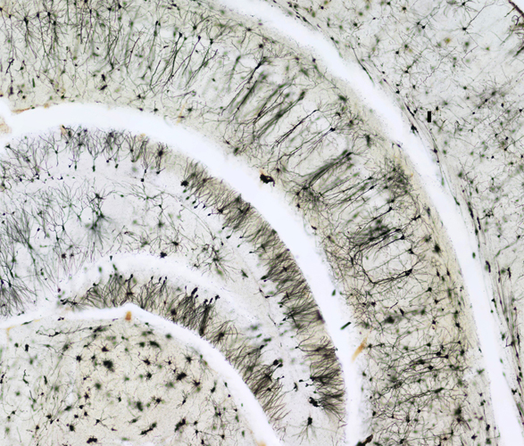
Specimen: Mouse brain, hippocampal region.
Technique: Brightfield.
Honourable Mention: Mrs. Magdalena Turzańska
University of Wrocław
Poland;
Specimen: Lepidozia reptans (pinnately branched leafy liverwort).
Technique: Autofluorescence with z-stack reconstruction, magnification 250x.
Honourable Mention: Dr. Andrew Woolley and Dr. Aaron Gilmour
University of New South Wales
Randwick, Australia;
Specimen: Complex cell culture based on mouse embryonic brain cells.
Technique: Confocal imaging, 200x.


Fantastic imagery... and proof that, when it comes to beauty and amazing symmetry, nothing beats nature. Love it. Thanks, TB.
ReplyDelete